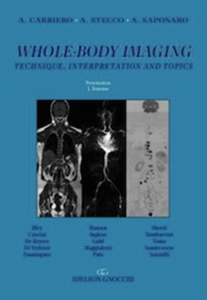
Whole-Body Imaging. Technique, interpretation and topics
85,00€ 80,75€
Formato 21×30 di pagine XVI-204 con 144 figure in 443 parti e 12 tabelle – Cartonato
Disponibile
Foreward
It is with great pleasure that I introduce Alessandro Carriero’s book “Whole-body imaging: technique,interpretation and topics”. Whole-body imaging is one of the most recent developments in the field of MRI and even though it is not yet commonly used in clinical practise it has intrinsically enormous potential thanks to its ability to add new functional information to traditional morphologic data. The book is divided into three sections that accurately deal with the physical basis, the different techniques and the main clinical applications inn adults and children. The chapters dedicated to its comparison with other imaging modalities like Fdg PET and low dose whole body CT are particularly interesting. The third section, an authentic gallery of clinical cases, concludes the book. My heartfelt congratulations to all those who contributed to the creation of this publication. I am certain it will be very useful to readers.
Lorenzo Bonomo
Introduction
Diffusion imaging is the closest tool, beetween diagnostic radiology and MRI, that have so far come to “functional” tissue imaging, and therefore also represents a sort of cultural bridgehead towards the functional methods of nuclear medicine. Tumours are the second most frequent cause of death in the developed world (the third most frequent in developing countries), accounting for 12.5% of all deaths, and the need for their diagnosis, staging and posttreatment follow-up makes diagnostic imaging a key element in the management of oncological patients. MR imaging based on the phenomenon of diffusion (DWI), which was originally developed for neuroradiological applications, has only recently been extended to the whole body because of the technical problems intrinsic to the cardinal sequences of DWI (echo planar imaging – EPI) and difficulties such as artefacts due to magnetic susceptibilitye and anatomical distorsions. The technical evolutions of MR coils and equipment, together with the introduction of parallel imaging, made it possible to overcome these problems and thus laid the foundations for the extension of DWI to the whole body. These developments have included the introduction of the “rolling table” and the use of increasingly more suitable sequences for the study of extra-cranial districts, which have also made it possible to obtain multiplanar reconstructions such as DWIBS. Atherosclerosis is a global problem and not a disease affecting just one sector. The increasing average life expectancy of patients is leading to the more frequent observation of the co-existence of complex problems that may affect different body’s districts but are often attributable to the same underlying pathology of atheromatous vascular disease. Further technical advances such as the use of “fresh blood” sequences make it possible to hypothesise the increasing application of study protocols to routine clinical practice. However, whole-body imaging is still in its pioneering phase and there are still many unanswered questions concerning its precise place in the process of clinical patient management. We believe that these questions will be answered by future multicentric trials, which will therefore be instrumental in offering radiologists a further cultural, as well as technical, evolution.
A. Carriero, A. Stecco, A. Saponaro
INDEX
Foreward
Introduction
SECTION I Whole-Bod y Imaging : from Techni que to Interpretation
1. Physical basis – MRI, DWI, MRA
2. Whole-body MRA
3. Whole-body MRI
4. Whole-body diffusion
SECTION II Topics
1. Whole-body MRI: Technique
2. Whole-body diffusion: Technique
3. Whole-body diffusion-weighted imaging with 3 Tesla: basic clinical concepts and advanced applications
4. Whole-body MRI: Normal, Pathologic and Pitfalls Experiences from the Population-Based SHIP-Study
5. Whole-body MRA: Normal, Pathologic and Pitfalls
6. Whole-body MRI and FDG-PET fused images for evaluation of patients with cancer: a pictorial essay
7. Whole-body MRI: approaches to tumor screening
8. Whole-body MRI for evaluation of bone metastasis in patient with malignant lymphoma
9. Multiple myeloma: Whole-body MRI vs. PET and Low Dose Whole-body CT
10. Whole-body Magnetic Resonance Imaging (WB-MRI) and bone scintigraphy to detect bone metastases: an update of UMG experience
11. Characterization in DWI and Whole-body diffusion
12. Whole-body MRA with contrast medium: diagnostic accuracy
13. A total atherosclerotic score for Whole-body MRA and its relation to traditional cardiovascular risk factors
14. Whole-body MRI in paediatrics
SECTION III CASES GALLERY
15. Clinical cases
16. Posters
PET-CT compared with Whole-body dwi /mri
Bone scan with Whole-body DWI/MRI: cases
Whole-body PET-MRI image fusion
Index
| Autori | |
|---|---|
| Editore | |
| Anno | |
| Lingua |
Prodotti correlati
Diagnostica per Immagini
Formato 15x21 di pagine X-116
Diagnostica per Immagini
DIAGNOSTICA PER IMMAGINI – RADIOLOGIA CONVENZIONALE – TAVOLE TEORICO-PRATICHE
Formato 30x21 di pagine XII-220 - Cartonato
Diagnostica per Immagini
Formato 21x30 di pagine XVI-688 con 1326 figure e 27 schemi - Cartonato
Diagnostica per Immagini
Formato 17x24 di pagine XII-124 con 71 figg. e 8 tabb.
Diagnostica per Immagini
Presentazione di L. Bonomo - Formato 21x30 di pagine XVI - 320 con 251 figure a colori e b/n e 10 tabelle - Cartonato
Diagnostica per Immagini
Formato 21x30 di pagine XXIV-672 e 1897 immagini a colori e b/n - Cartonato
Diagnostica per Immagini
Formato 17x24 di pagine VII-212 con 208 figg. e 18 tabb.
Diagnostica per Immagini
Formato 21x30 di pagine XVIII-206 con 294 figure e 34 tabelle - Cartonato
Diagnostica per Immagini
V Edizione Formato 21x30 di pagine XXIV–1192 con 1071 figure a colori e b/n e 78 tabelle - Cartonato con sopracoperta
Anatomia
Formato 30x21 di pagine XX-236 con 239 figg. a colore e B/N - Cartonato
Diagnostica per Immagini
a cura di A. Ragozzino - Formato 20x27 di pp. XX-416 con 743 figure a colori e b/n
Diagnostica per Immagini
Tre volumi indivisibili Formato 21x30 di complessive pagine LXXXVIII-1956 con 3186 figg. a colori e b/n e 121 tabelle - Cartonati in cofanetto

